# Insights on Cytoplasmic Pigments in Human Cells: A Comprehensive Overview
Written on
Chapter 1: Understanding Pigments in Human Cells
Pigments are specific compounds responsible for reflecting certain wavelengths of light, which results in color. In human cells, pigment granules—membrane-enclosed vesicles containing pigments or their precursors—can accumulate as part of metabolic processes (i.e., as waste) or serve particular functions, such as protection against UV radiation. The primary pigments found in human cytoplasm are melanin, lipofuscin, and ceroid pigments. The latter two share similarities and are often linked to aging. This is distinct from chromatophores, like retinal and rhodopsin in the eyes, and porphyrins (such as heme) found in bodily fluids.
Section 1.1: Melanin - The Protective Pigment
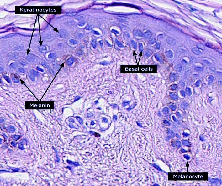
Melanin is essential for providing color to skin, hair, and eyes while also offering protection from ultraviolet light (e.g., sunlight). This pigment is synthesized by specialized cells known as melanocytes, as well as retinal pigment epithelial cells in the eyes. Melanocytes possess organelles called melanosomes, which transfer melanin to keratinocytes, contributing to pigmentation in hair and skin.

Exposure to UV light triggers the production of eumelanin, one of two melanin types in mammals (the other being pheomelanin). Eumelanin appears brown to black, while pheomelanin is blond to red. Notably, melanin is also present in the brain. The substantia nigra, a brain region rich in neurons, derives its name from the black pigment found within its cells, which serves as a byproduct of dopamine production. DOPA (dihydroxyphenylalanine), a precursor of melanin, is also present in the brain. The loss of melanin-rich cells in the substantia nigra is associated with Parkinson’s Disease. Changes in melanin levels due to age or disease can lead to grey hair, age spots, and even skin cancer (melanoma). For further reading on